- This topic is empty.
-
AuthorPosts
-
19/11/2026 at 17:53 #86163
■ 1. Background
AP-III-a4 (T6759), CAS: 1177827-73-4 is a small-molecule enolase inhibitor distinguished as the first nonsubstrate analogue reported for this glycolytic enzyme, with an IC50 of 0.576 μM. Enolase catalyzes the dehydration of 2-phosphoglycerate to phosphoenolpyruvate (PEP), a rate-contributing step near the terminus of the Embden–Meyerhof pathway that feeds substrate-level phosphorylation at pyruvate kinase. By inhibiting enolase, AP-III-a4 acutely lowers PEP levels and constrains glycolytic flux, thereby diminishing ATP yield derived from glycolysis and altering redox balance coupled to lactate production. The nonsubstrate analogue nature implies engagement of an interaction surface distinct from the 2-phosphoglycerate/PEP mimicry typical of classical enolase inhibitors, a feature useful for dissecting active-site versus allosteric control of enolase catalysis and for probing isoform-agnostic versus isoform-biased target engagement across ENO1/ENO2/ENO3 in different cell types [1].
Within a pathway framework, glucokinase initiates the glucose-utilization cascade by producing glucose-6-phosphate, setting the upper boundary of glycolytic input, whereas enolase acts downstream to shape the throughput to pyruvate and lactate. AP-III-a4 thus enables experiments that decouple input control (glucokinase) from distal catalytic constraints (enolase), allowing researchers to quantify how distal bottlenecks propagate to whole-pathway flux control, metabolite pooling, and energy charge. Under conditions where cells rely heavily on glycolysis to sustain ATP and biosynthetic precursors, enolase inhibition by AP-III-a4 imposes metabolic stress that can converge on intrinsic apoptotic signaling. Canonical components of this coupling include AMP/ATP-sensitive sensors and mitochondrial effectors (e.g., BCL-2 family control of outer membrane permeabilization), culminating in caspase activation when energetic and redox thresholds are breached [2]. Such cross-talk between metabolism and the apoptosis machinery is central to understanding how glycolytic nodes govern cell survival decisions.
AP-III-a4 is therefore a versatile probe for: mapping enolase’s control coefficient within the glucokinase-to-pyruvate axis using time-resolved metabolomics and 13C-flux analysis; benchmarking glycolysis-dependent apoptotic checkpoints by correlating PEP, ATP, and NAD(H) dynamics with caspase activity; and differentiating enolase-specific effects from upstream perturbations through comparative studies with genetic modulation of glucokinase or phosphofructokinase. Its submicromolar potency facilitates acute, titratable pathway modulation compatible with live-cell imaging, extracellular flux assays (oxygen consumption and acidification), and lactate/pyruvate secretion profiling. Because enolase also exhibits context-dependent “moonlighting” roles at the cell surface and in RNA metabolism, AP-III-a4 provides an entry point to test how catalytic inhibition intersects with these functions without confounding substrate mimicry [1]. Collectively, AP-III-a4 is well suited for mechanistic dissection of glycolytic control, for delineating the link between carbohydrate metabolism and apoptosis, and for building quantitative models that connect glucokinase-driven input with distal enzymatic constraints and fate-determining signaling outputs [2].
■ 2. Literature review
■ 2.1 KBTBD11 suppresses hepatocellular carcinoma by targeting ENO1-mediated glycolysis.
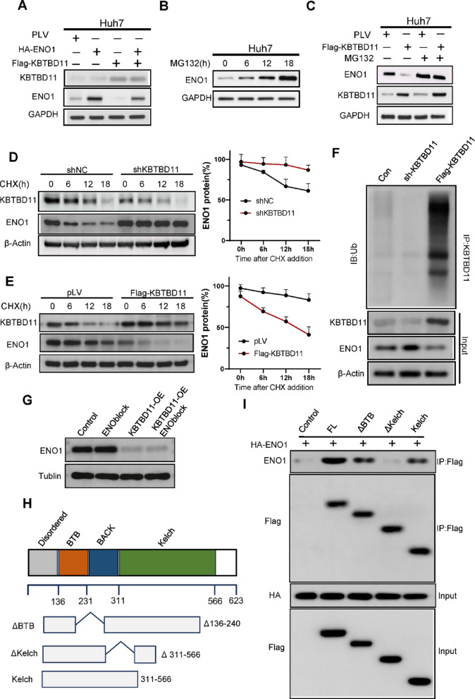
▲ KBTBD11 promotes ENO1 degradation through the ubiquitin–proteasome pathway. (A) Western blot analysis of ENO1 protein levels in Huh7 cells transfected with control vector, HA-ENO1, and/or Flag-KBTBD11. (B) Time-course analysis of ENO1 protein accumulation following treatment with the proteasome inhibitor MG132 (15 nM) for the indicated durations in Huh7 cells. (C) Immunoblot showing that MG132 (15 nM, 12 h) treatment reverses the KBTBD11-induced downregulation of ENO1 protein in Huh7 cells. (D–F) Cycloheximide (CHX) chase assays were performed to assess ENO1 protein stability after KBTBD11 knockdown (shKBTBD11), overexpression (Flag-KBTBD11), or control. Huh7 cells were treated with CHX (20 µg/mL) for the indicated time points. (E) Co-immunoprecipitation of ENO1 followed by immunoblotting with an anti-ubiquitin antibody to assess ubiquitination levels in Huh7 cells transfected with or without Flag-KBTBD11. MG132 (15 nM, 12 h) was used to stabilize ubiquitinated proteins prior to lysis. (F) Ubiquitination of ENO1 in Huh7 cells with KBTBD11 overexpression or knockdown, assessed by immunoprecipitation followed by Western blot. (G) Western blot analysis of ENO1 expression in Huh7 cells treated with AP-III-a4 (ENOblock) alone or in combination with KBTBD11 overexpression. (H) Schematic of the KBTBD11 domain architecture and deletion constructs. (I) Co-immunoprecipitation analysis of the KBTBD11–ENO1 interaction in 293T cells using full-length and domain-deletion mutants of KBTBD11.
AP-III-a4 (T6759) was described as a pharmacological inhibitor of ENO1, and its administration in hepatocellular carcinoma models recapitulated the metabolic suppression seen with KBTBD11 overexpression. In this study, AP-III-a4 was tested in Huh7 and Hepa1-6 cells, where investigators measured extracellular acidification rate, ATP, and lactate to evaluate glycolytic activity after intervention. AP-III-a4 reproduced the suppression of these metabolic outputs that was observed when KBTBD11 was overexpressed, indicating that direct pharmacological inhibition of ENO1 can mimic the metabolic effects linked to reduced glycolytic flux. Beyond metabolic readouts, AP-III-a4 also affected cellular proliferation: AP-III-a4 treatment significantly impaired growth in both Huh7 and Hepa1-6 cells. When combined with KBTBD11 overexpression, AP-III-a4 produced a further reduction in cell viability, supporting that pharmacologic ENO1 inhibition and KBTBD11 converge functionally on glycolytic control to limit tumor cell growth. Collectively, these findings position AP-III-a4 as an ENO1-targeting agent that suppresses glycolysis-associated metabolic outputs and reduces viability in hepatocellular carcinoma cell lines, with enhanced effects on viability when applied alongside KBTBD11 overexpression.[3]
■ 2.2 LncPRESS1 promotes the progression and cisplatin resistance of breast cancer through upregulation of ENO1 and activation of glycolysis.
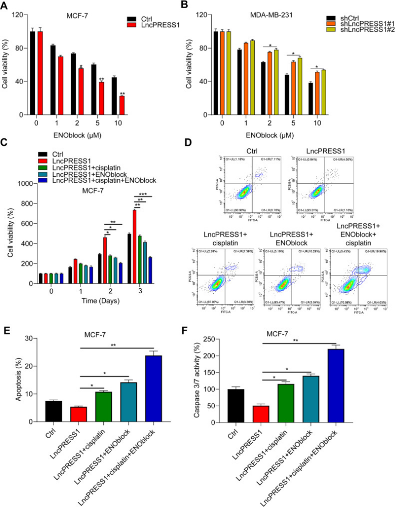
▲ ENO1 inhibitor promotes the sensitivity of cisplatin in BRCA cells. (A, B) MDA-MB-231 and MCF-7 cells with knockdown or overexpression of LncPRESS1 were treated with different concentrations (0, 1, 2, 5, 10 µM) of AP-III-a4 (ENOblock) for 48 h. Cell viability was assessed by CCK-8 assay, and each group was normalized to the 0 µM group. *p < 0.05. (C) Ctrl and LncPRESS1-overexpressing MCF-7 cells were treated with vehicle, AP-III-a4 (ENOblock, 2 µM), cisplatin (20 µM), or their combination for different times, and cell viability was measured by CCK-8 assay. *p < 0.05, **p < 0.01, ***p < 0.001. (D, E) Ctrl and LncPRESS1-overexpressing MCF-7 cells treated with vehicle, AP-III-a4 (ENOblock, 2 µM), cisplatin (20 µM), or their combination for 24 h were subjected to apoptosis analysis using PI/Annexin V-FITC staining on a flow cytometry system. *p < 0.05, **p < 0.01. (F) The activity of caspase 3/7 was examined using an assay kit in Ctrl and LncPRESS1-overexpressing MCF-7 cells treated with vehicle, AP-III-a4 (ENOblock, 2 µM), cisplatin (20 µM), or their combination for 24 h. *p < 0.05, **p < 0.01. The results demonstrate that AP-III-a4 (ENOblock) significantly enhances cisplatin-induced apoptosis and caspase 3/7 activation, particularly in LncPRESS1-overexpressing cells.
AP-III-a4 (T6759) was investigated as an ENO1 inhibitor in breast cancer cells and showed activity modulated by LncPRESS1 status. Overexpression of LncPRESS1 promoted the cytotoxicity of AP-III-a4, whereas LncPRESS1 knockdown suppressed cellular sensitivity to AP-III-a4 in breast cancer models. In functional combination studies, AP-III-a4 together with cisplatin dramatically inhibited the growth of MCF-7 cells, indicating a synergistic interaction between the two agents. Mechanistically linked readouts demonstrated that concurrent administration of AP-III-a4 and cisplatin significantly enhanced apoptosis and elevated caspase 3/7 activity, supporting potentiation of pro-apoptotic signaling under combination conditions. These effects were further contextualized by dosing parameters: MDA-MB-231 and MCF-7 cells engineered for LncPRESS1 overexpression or knockdown were exposed to 0–10 µM AP-III-a4 for 48 hours for viability assays, and fixed-dose combination experiments in MCF-7 cells used 2 µM AP-III-a4 and 20 µM cisplatin. Importantly, in the setting of LncPRESS1 overexpression—a context associated with reduced response to cisplatin—the application of AP-III-a4 overcame cisplatin resistance. Together, the study provides direct experimental evidence that AP-III-a4 exerts cytotoxic effects that are enhanced by LncPRESS1 overexpression, synergizes with cisplatin to suppress breast cancer cell growth, augments apoptosis and caspase activity under combination conditions, and restores cisplatin sensitivity in LncPRESS1-overexpressing cells.[4]
■ 2.3 Targeting Kupffer Cell Enolase 1 Attenuates Liver Inflammation and Injury in Hemorrhagic Shock.
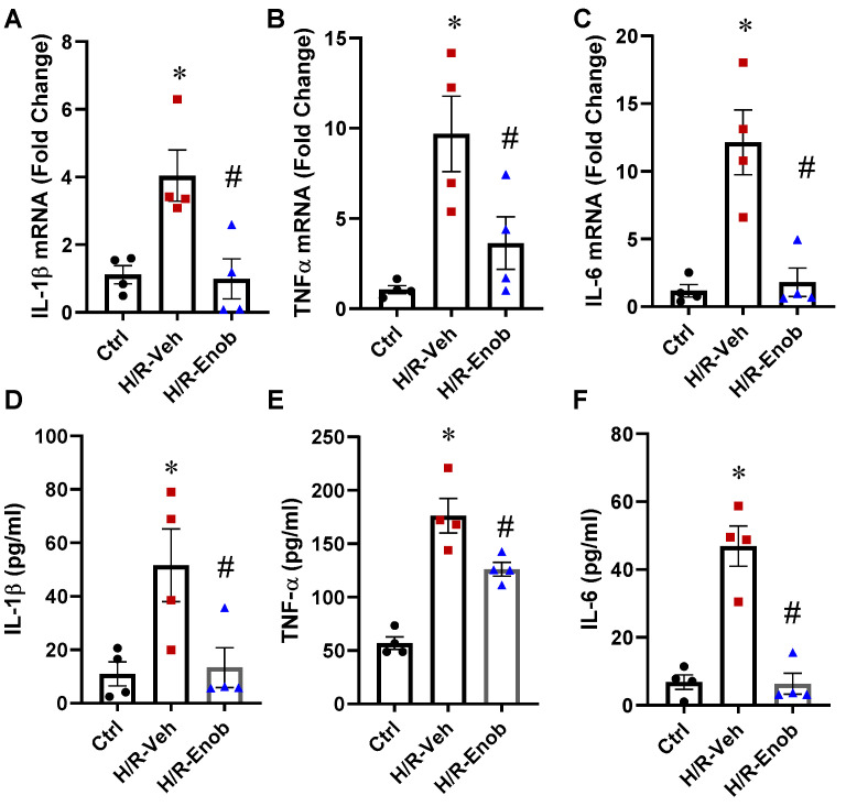
▲ ENOblock (AP-III-a4) alleviates H/R-induced Kupffer cell inflammation. Kupffer cells (KCs) were subjected to H/R, after which cells and the supernatant were harvested. Total RNA was extracted to assess the mRNA expression of IL-1β (A), TNF-α (B), and IL-6 (C) by qPCR. The supernatant was collected for cytokine analysis, and the levels of IL-1β (D), TNF-α (E), and IL-6 (F) were measured using ELISA kits. The results showed that H/R significantly upregulated the mRNA and protein expression of these pro-inflammatory cytokines, whereas ENOblock (AP-III-a4) treatment significantly suppressed these elevations. Data are presented as mean ± SEM (n = 4–6/group) and were analyzed using ANOVA and the SNK test. * p < 0.05 vs. Sham; # p < 0.05 vs. H/R-Veh.
AP-III-a4 (T6759) is described as a specific ENO1 inhibitor that suppresses glycolytic activity. In a hemorrhagic shock model, AP-III-a4 markedly dampened hepatic inflammation: it significantly mitigated HS-induced mRNA increases of IL-1β, TNF-α, and IL-6 by 78%, 83%, and 77%, and reduced the chemokines MIP-2 and KC by 78% and 49%. Consistent with targeting ENO1, AP-III-a4 decreased ENO1 enzymatic activity in Kupffer cells by 32% after hemorrhagic shock. Histologically, AP-III-a4 attenuated liver tissue alterations associated with hemorrhagic shock and lowered the Suzuki liver injury score by 42%. It also reduced hepatocyte apoptosis, as indicated by a 61% decrease in TUNEL-positive cells. At the cellular signaling level, AP-III-a4 lessened inflammasome-related activation in Kupffer cells, reducing both the frequency and mean fluorescence intensity of c-caspase-1–positive cells by 44% and 15%, and decreasing c-caspase-1 protein levels by 43%. In an in vitro hypoxia/reoxygenation setting with Kupffer cells, AP-III-a4 significantly decreased the pro-inflammatory cytokines IL-1β, TNF-α, and IL-6. Together, these findings show that AP-III-a4 dampens inflammatory cytokine and chemokine expression, reduces ENO1 activity in hepatic macrophages, alleviates histological liver injury, and limits apoptosis and c-caspase-1 activation following hemorrhagic shock and in hypoxia/reoxygenation-activated Kupffer cells.[5]
■ 2.4 Paeoniflorin Directly Targets ENO1 to Inhibit M1 Polarization of Microglia/Macrophages and Ameliorates EAE Disease.
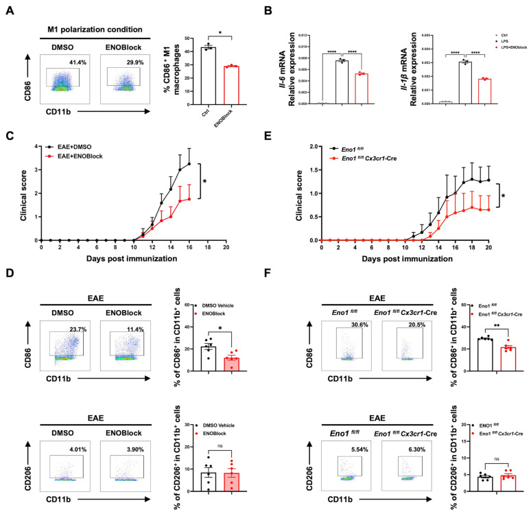
▲ ENOBlock (AP-III-a4) treatment or specific knockout of Eno1 in microglia inhibits M1 polarization and alleviates EAE progression. (A, B) BMDMs were treated with ENOBlock (AP-III-a4) (20 μM) in M1 polarization condition (LPS 100 ng/mL) for 24 h (n = 3). The percentage of CD86+ M1 BMDMs was analyzed by flow cytometry (A), and the mRNA expression of *Il-6* and Il-1β in M1 BMDMs was analyzed by qPCR (B). (C) The clinical score of MOG-induced EAE mice treated with DMSO vehicle or ENOBlock (AP-III-a4) (5 mg/kg) intraperitoneally from day 5 post immunization (n = 6). (D) Flow cytometry analysis of CNS mononuclear cells isolated from DMSO vehicle and ENOBlock-treated mice on day 16 post immunization. Representative data and percentages of CD86+ or CD206+ cells in CD11b+ cells in the CNS from the two groups (n = 6). (E) The clinical score of MOG-induced Eno1 fl/fl and Eno1 fl/fl Cx3cr1-Cre EAE mice (n = 6). (F) CNS mononuclear cells isolated from Eno1 fl/fl and Eno1 fl/fl Cx3cr1-Cre EAE group mice on day 16 post immunization were analyzed by flow cytometry. Representative data and percentages of CD86+ or CD206+ cells in the CNS from the two groups (n = 6). Data are presented as mean ± SEM; * p < 0.05, ** p < 0.01, **** p < 0.0001; ns, no significance.
AP-III-a4 (T6759) was evaluated for its impact on inflammatory polarization and disease severity in experimental autoimmune encephalomyelitis (EAE) and in vitro macrophage assays. In LPS-stimulated bone marrow–derived macrophages, AP-III-a4 reduced M1-associated features, as evidenced by a decrease in the percentage of CD86+ cells and a reduction in mRNA levels of the M1-type cytokines Il-6 and Il-1β. In the in vivo EAE model, administration of 5 mg/kg AP-III-a4 alleviated clinical severity. Correspondingly, AP-III-a4 reduced the percentage of CD86+ CD11b+ M1 microglia/macrophages compared with the vehicle group. Together, these findings indicate that AP-III-a4 suppresses markers of M1 polarization in macrophages in vitro and decreases M1 microglia/macrophage abundance in vivo, coinciding with an improvement in disease severity in EAE. The evidence supports AP-III-a4’s capacity to downshift proinflammatory signatures across cellular and organismal contexts relevant to neuroinflammation, linking reduced M1 polarization with ameliorated clinical manifestations in the EAE model.[6]
■ 3. Conclusion
AP-III-a4 functions as a small-molecule enolase inhibitor that noncompetitively suppresses the catalytic conversion of 2-phosphoglycerate to phosphoenolpyruvate, thereby acutely lowering PEP levels and constraining glycolytic flux. Literature findings demonstrate that AP-III-a4 effectively inhibits ENO1-mediated glycolysis, reduces metabolic outputs such as ATP and lactate, and impairs cancer cell proliferation in hepatocellular carcinoma and breast cancer models. It synergizes with genetic and chemotherapeutic interventions, overcoming drug resistance and promoting apoptosis. Additionally, AP-III-a4 attenuates liver inflammation and injury by suppressing ENO1 activity in Kupffer cells and reduces neuroinflammatory M1 polarization in macrophages, improving disease outcomes in experimental autoimmune encephalomyelitis. Future research could explore AP-III-a4’s mechanistic roles in linking metabolism to apoptosis, dissect isoform-specific effects in diverse cell types, and evaluate therapeutic potential in metabolic, inflammatory, and cancer conditions.
■ 4. Q&A
Q1: What is the mechanism of action of AP-III-a4 on glycolysis?
A1: AP-III-a4 noncompetitively inhibits enolase, blocking the conversion of 2-phosphoglycerate to phosphoenolpyruvate (PEP), which lowers PEP levels, constrains glycolytic flux, reduces ATP production, and alters redox balance linked to lactate formation.
Q2: How does AP-III-a4 affect cancer cell viability and proliferation?
A2: AP-III-a4 inhibits ENO1-mediated glycolysis in hepatocellular carcinoma and breast cancer cells, reducing metabolic outputs and cell growth. It enhances apoptotic signaling and shows synergy with KBTBD11 overexpression and cisplatin treatment, overcoming drug resistance and further decreasing viability.
Q3: What are the anti-inflammatory effects of AP-III-a4 described in the literature?
A3: AP-III-a4 suppresses ENO1 activity in Kupffer cells, reduces pro-inflammatory cytokines and chemokines (IL-1β, TNF-α, IL-6, MIP-2, KC), limits inflammasome activation and caspase-1 cleavage, thereby attenuating liver inflammation and injury in hemorrhagic shock models. It also inhibits M1 polarization of microglia/macrophages, reducing neuroinflammation and disease severity in experimental autoimmune encephalomyelitis.
■ 5. Reference
[1] Pancholi V. Multifunctional alpha-enolase: its role in diseases. Cell Mol Life Sci. 2001;58(7):902-20.
[2] Vander Heiden MG, Cantley LC, Thompson CB. Understanding the Warburg effect: the metabolic requirements of cell proliferation. Science. 2009;324(5930):1029-33.
[3] Liu Y, Chen Y, Tang L, Lin F, Zhuang Q, Chen H, et al.. KBTBD11 suppresses hepatocellular carcinoma by targeting ENO1-mediated glycolysis.. Journal of translational medicine. 2025;23(1):1087.
[4] Ke L, Gao W, Leng Y, Chen J, Chen H, Li S, et al.. LncPRESS1 promotes the progression and cisplatin resistance of breast cancer through upregulation of ENO1 and activation of glycolysis.. Cell division. 2025;20(1):23.
[5] Hu Z, Li J, Rashid N, Jacob A, Wang P. Targeting Kupffer Cell Enolase 1 Attenuates Liver Inflammation and Injury in Hemorrhagic Shock.. International journal of molecular sciences. 2025;26(17):.
[6] Sun Y, Wang G, Li S, Jiang Y, Liu Y, Gao Y, et al.. Paeoniflorin Directly Targets ENO1 to Inhibit M1 Polarization of Microglia/Macrophages and Ameliorates EAE Disease.. International journal of molecular sciences. 2025;26(8):.
http://www.targetmol.com
TargetMol Chemicals Inc. -
AuthorPosts

- You must be logged in to reply to this topic.